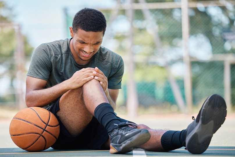
Knee Injuries

Knee injuries can be broken down into acute/traumatic injuries and non acute overuse injuries.
Acute Knee
Signs that you might have a significant acute knee injury warranting physio assessment include:
- Swelling
- Instability / giving way
- Locking (where the knee gets stuck and cannot be moved)
- A popping sound at the time of injury
One of the most common traumatic injuries we see is the ACL rupture. The ACL connects the thigh to the shin bone and helps keep your knee stable when pivoting and changing direction. The ACL is usually injured when the knee buckles excessively inwards whilst an athlete is attempting to change direction. An ‘pop’ is usually heard and the knee will then swell. This is often accompanied by an intense pain for a few minutes following the injury which then subsides. Sometimes the athlete may then believe he is then able to continue playing. An uneasiness and instability the ensues. Sports that often see this injury include netball, all football codes, basketball and skiing.
The decision to have surgery after an ACL injury depends upon the patient’s age and activity level. Usually younger active individuals will choose to undertake surgical reconstruction of the ACL. Whereas older sedentary persons may try conservative treatment consisting of strength and ROM exs. We have seen successful outcomes with conservative treatment alone. Over the years we have successfully rehabbed hundreds of ACL repairs. Our rehab protocols have continued to improve over time and our rate of re-rupture has been extremely low.
Medial collateral ligament (MCL) tears can also occur when the knee buckles inwards. Pain and swelling is focused on the inner aspect of the knee joint. There is often a stiffness or pulling at the extremes of flexion and extension. Isolated MCL injuries will usually have good results with physiotherapy rehab. An MRI scan is useful to grade the injury. Grade 1 sprains can be managed with taping to stabilise the knee for 1-2 weeks. For Grade 2 and 3 MCL tears it is important to wear a ROM brace to keep the pressure of the ligament as it heals. Crutches can be required for the first couple of weeks for more severe MCL tears. Treatment also should comprise of massage to reduce stiffness and scar tissue adherence and a progressive ROM/strengthening program. Full recovery can take from 3-4 weeks to 12 weeks depending on the grade of tear.
Patella dislocation occurs when the knee cap (patella) 'pops' out of place. A knock to the side of the knee can often cause this. On other occasions an individual's anatomy may predispose them to a patella dislocation and a simple twistintg motion on the knee can cause the injury. Sometimes the injury will be obvious as the patient will see the patella sitting to the outside of the knee and medical assistance will be required to relocate it. In other instances the patella will dislocate and then relocate quickly and the injury will be more vague to the patient. A patella dislocation can present similarly to an ACL injury with swelling and pain around the front of the knee. Physical examination usually reveals an apprehension when the patella is moved to the side. First time dislocations are nearly always treated with physiotherapy rehab. A brace is used to keep the knee straight for several weeks whilst the ligaments heal and then ROM and strength gradually restored. Persons who experience multiple dislocations can consider a reconstruction which in our experience has good results.
Overuse or chronic knee injuries will generally affect either the tendons or joint cartilage.
The degenerative knee
Cartilage lines the ends of the bones that form the knee joint. It is an extremely durable tissue and it acts as a shock absorber. As you enter into your 40s, 50s and 60s the knee may begin to show signs of wear and tear as the cartilage and meniscus begin to thin, tear or degenerate. Pain, joint swelling, stiffness, grinding, clicking and catching can all be symptoms of this process. This is all synonymous with osteoarthritis. There are varying degrees of osteoarthritis and it is important not to panic if you have been diagnosed with it. Most people over the age of 40 will have arthritic signs on MRIs and x-rays and many of these people are pain free. In some ways it is akin to getting some grey hairs as you age.
We have seen many people with early-moderate arthritic symptoms stay on top of their pain by following a few of our recommendations. New evidence is highlighting the fact that arthoscopy is frequently no better than placebo for arthritic knees. Many people will find benefit from loosing weight and strengthening the muscles around the painful joint. It is important to keep a positive attitude about the injury as many people will avoid movement out of fear and this sedentary behaviour can lead to increased pain. We like to adopt the adage of 'motion is lotion'. It is important to understand that your body is not a machine that can wear out. In contrast it is a living organism that depends upon movement to enhance blood flow to maintain tissue health. Specific exercise at an appropriate intensity is important for persons with a degenerative knee. If you do too much too soon before your body can adapt this can promote injury. We have seen many examples of people who have maintained a healthy body weight and lifestyle keep their arthritic knee in check.
Complications
One of the main complications we see after a knee injury or surgery is the development of a stiff knee where it is extremely difficult to bend and/or straighten the knee. This is due to excessive scar tissue formation around the knee joint and this process is referred to as arthrofibrosis. It is usual to expect some stiffness after even minor arthroscopic procedures but this usually passes with time and range of motion exercises. With arthrofibrosis the stiffness can be extremely stubborn. We have found that low load sustained stretches are the most effective intervention for this issue and depending on the severity of stiffness we usually find a particular position to allow for a long stretch into the restricted motion. This exercise works on the ‘creep’ process to gain improvements in the scar tissue extensibility. If possible, cycling, even using an upper body ergometer for the legs or bike are also extremely beneficial. Whilst patients often focus on knee bending, a deficit of extension or inability to fully straighten the knee can be more troublesome as it will affect gait significantly. It is extremely important to identify this issue as early as possible.